The radiographic analysis/ x-ray analysis is still one of the most important instruments in the dental diagnosis.
With digital radiography the precision of the radiographs noticeably has improved, while the exposure levels have been reduced to a tolerable level at the same time. Such x-rays give the dentist a clear picture of structures in the mouth, that are otherwise not visible, especially in the bone and cartilage, as well as the positions and dimensions of neighbouring structures, such as nerve canals, sinuses and jaw joints.
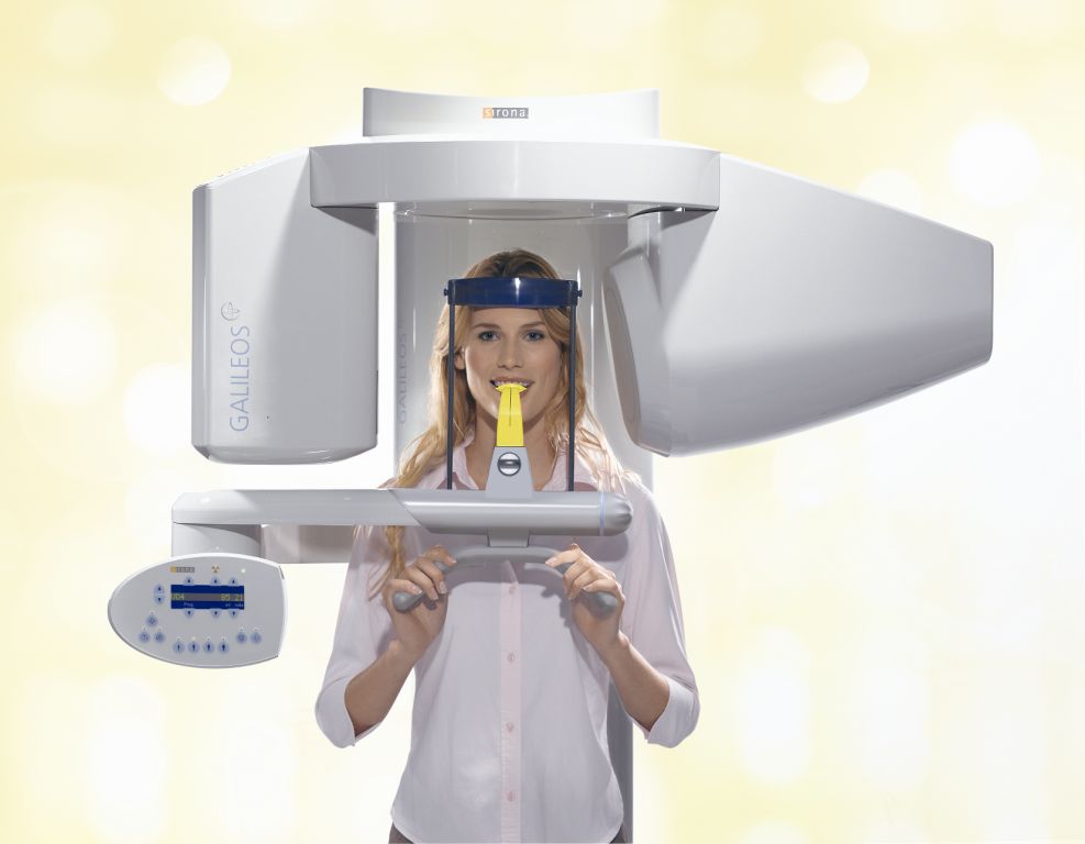
New technology makes it possible to produce 3D pictures that display the real life situation much better and more realistically. This is especially important during the planning for implant placement, but can also give vital information in periodontal disease and root canal treatment.
Digital radiography
We use digital radiography for single tooth, Panoramic radiographs and 3D pictures in our surgery. This enables us to provide you with faster diagnosis, better communication with our referring colleagues and most of all the reduction of exposure levels of up to 90% for our patients.
There are no more environmentally harmful development chemicals that have to be disposed off. A special sensor is used instead of a film. This sensor does not have to be developed anymore, but converts the x-rays into digital signals.
Those are processed by the computer then, so that the picture can be seen on the monitor immediately. This enables the dentist to analyse it straight away and support a diagnosis.